Stanford Microscopy Facility
OVERVIEW
MICROSCOPY.STANFORD.EDU RANKINGS

Date Range

Date Range

Date Range
LINKS TO WEB SITE
We study the primary cilium, a surface-exposed organelle required for vision, olfaction and developmental signaling and whose dysfunction leads to. 8203; obesity, skeletal malformations and kidney cysts. To decode the fundamental principles of ciliary trafficking and to understand. How trafficking shapes signaling at the primary cilium, we leverage. A broad expertise in biochemistry, proteomics, cell biology and in vitro reconstitution.
WHAT DOES MICROSCOPY.STANFORD.EDU LOOK LIKE?
MICROSCOPY.STANFORD.EDU HOST
BROWSER ICON

SERVER OS
I detected that this website is weilding the Apache server.PAGE TITLE
Stanford Microscopy FacilityDESCRIPTION
The Cell Sciences Imaging Facility CSIF is a University service center that provides high resolution, state-of-the-art light and electron microscopy technologies for imaging and analyzing the molecular and structural organization of cells, tissue and bioengineered materials.CONTENT
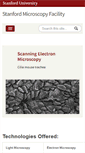
This site microscopy.stanford.edu states the following, "Access for non-Stanford, external users." We noticed that the website said " Eva Huang, Dunn Lab, human embryonic stem cells." It also stated " Confocal microscopy scanning and spinning-disk. Transmitted-light imaging phase, DIC, histology. High-content screening Confocal and Wide-field. Super-resolution imaging STORM, STED, SIM and AiryScan. Cell surface imaging with 100 nM z-resolution TIRF. Specialized microscopy FRAP, FRET, FCS, FLIP." The header had confocal as the highest ranking keyword. It was followed by csif, Cell Sciences Imaging Facility, and electron microscopy which isn't as ranked as highly as confocal. The next words the site used was fluorescence. light microscopy was also included but could not be viewed by search engines.SEEK MORE DOMAINS
We are promoting cutting edge research in basic and applied sciences through research and development activities, as well as quality training and education. Through individual training, short courses and formal courses that can be taken for credit. Click on the image to learn more. Selected labs in the MI.
Invasive hyphal growth of Magnaporthe oryzae in live rice cells. oryzae were incubated on the rice sheath.
Arkansas Nano and Bio Materials Characterization Facility. Nanoscale instruments and expertise for campus and the state. 2015, The University of Arkansas.
Please note that links from this site point to an updated Microscopy Core Website. The UCSD Health Sciences Microscopy Core is a state-of-the-art imaging core facility that serves the needs of laboratories in and outside of the UCSD School of Medicine. The Core strives to promote interdisciplinary, collaborative research among the local research community.
Darr; Skip to Main Content. CoolLED pE - 300 lite. CoolLED pE - 300 ultra. Two Chambered McMaster slide with imprinted grid. Three Chambered McMaster slide with imprinted grid.